
Dental question about observed-topic:dark-gums
Dark gum tissue can be a normal melanin pattern or a sign of irritation, medication effect, inflammation, or a lesion that needs examination.
The short answer
Dark gums are not one diagnosis. Healthy gingiva may contain more melanin, while new or uneven pigmentation can follow irritation, smoking, medication exposure, inflammation, or a pigmented oral lesion.
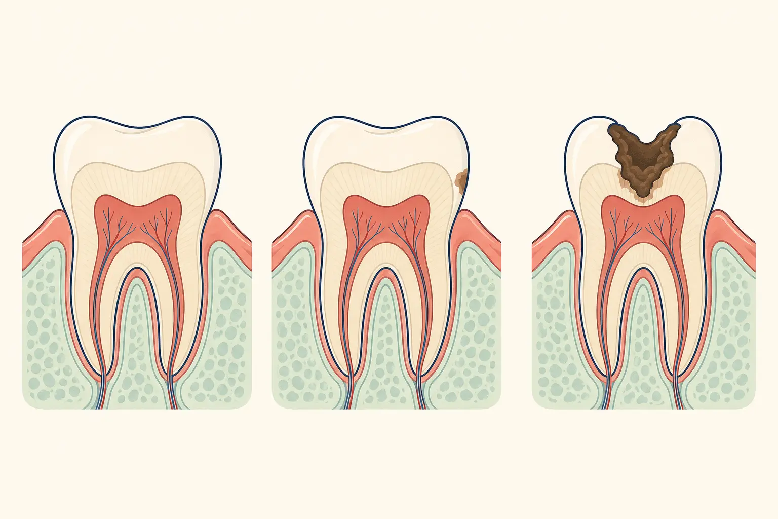
What is the direct answer?
What does this question mean?
Gum color comes from melanin in the epithelial layer, with blood supply and thickness affecting its appearance. A clinician considers whether the change is flat or raised, uniform or irregular, and stable or progressive.
What can you do next?
Take clear photographs in consistent lighting and note when the change began, any new medicine, tobacco exposure, bleeding, pain, or trauma. Do not scrub or use bleaching products on the gums.
When should you seek dental care?
Arrange an examination for a new, enlarging, irregular, bleeding, ulcerated, or one-sided dark area. The dentist may inspect the mouth, review medicines and habits, photograph the site, and refer for biopsy when indicated.
What should you ask the dentist?
Ask whether the pattern looks physiologic or acquired, what changes should be monitored, and whether periodontal treatment, medication review, or specialist assessment is appropriate.